
IMPACTED CANINES
Understanding Impacted Canines & Specialized Care
Impacted canines are teeth that do not properly erupt into their normal position in the mouth and remain partially or completely trapped beneath the gums or jawbone. The upper canine teeth, also called cuspids, play an important role in your bite, jaw function, and smile appearance. When these teeth fail to erupt correctly, they can affect surrounding teeth, create alignment problems, and increase the risk of future oral health complications. Oral and maxillofacial surgeons specialize in diagnosing and treating impacted canines using advanced surgical techniques designed to preserve the natural tooth whenever appropriate.
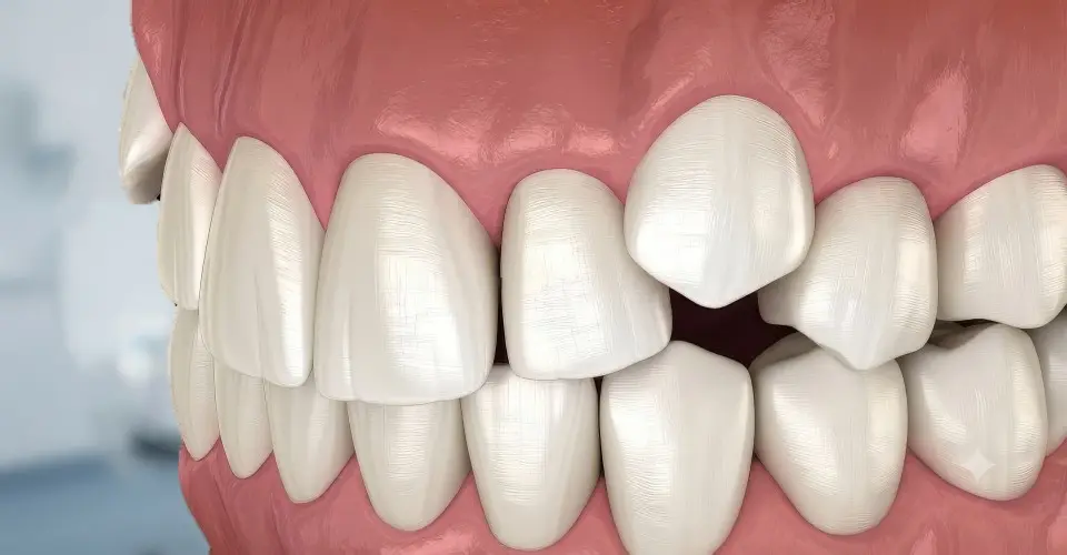
Importance of Canines & Early Diagnosis
Canines are among the most important teeth in the mouth because they help guide the movement of the jaw during chewing and contribute significantly to the appearance and function of the smile. Unlike wisdom teeth, which are often removed when impacted, impacted canines are usually preserved whenever possible because of their importance in maintaining a healthy bite. Treatment often involves a coordinated approach between an oral surgeon and orthodontist to help guide the tooth into its proper position.
Impacted canines most commonly occur in the upper jaw and are often discovered during routine dental examinations or orthodontic evaluations. In many cases, there are no obvious symptoms, and the condition is identified through dental X-rays or advanced 3D imaging. Early diagnosis is important because timely treatment may improve the chances of successfully guiding the tooth into alignment and preventing damage to nearby teeth.
Causes & 3D CBCT Evaluation
There are several reasons why a canine tooth may become impacted. Common contributing factors include limited space in the dental arch, abnormal tooth development, overcrowding, retained baby teeth, unusual growth patterns, or the presence of cysts or other obstructions preventing normal eruption. In some cases, the canine may develop in an abnormal direction and become positioned against the roots of neighboring teeth, potentially causing damage if left untreated.
Your oral and maxillofacial surgeon will perform a detailed evaluation to determine the exact location and position of the impacted canine. Advanced imaging, including 3D cone beam computed tomography (CBCT), provides a detailed view of the tooth, surrounding bone, nearby tooth roots, and important anatomical structures. This information allows your surgical team to create a precise treatment plan while helping minimize risks and improve the likelihood of a successful outcome.
Surgical Exposure, Orthodontic Coordination & Comfort
Treatment for impacted canines typically involves a combination of orthodontic and surgical care. In many cases, an orthodontist first creates enough space in the dental arch for the canine to move into position. The oral surgeon then performs a procedure called surgical exposure, where the gum tissue covering the impacted tooth is carefully opened to allow access to the tooth. A small orthodontic attachment may be placed on the tooth, allowing the orthodontist to gradually guide it into its proper location.
The surgical exposure procedure is typically performed using local anesthesia, with sedation and anesthesia options available for patients who experience dental anxiety or require additional comfort. During the procedure, your oral surgeon carefully accesses the impacted tooth while preserving surrounding tissues and structures. The goal is to provide the orthodontist with the access needed to safely and gradually move the tooth into alignment.
Alternative Options, Complications, Recovery & Collaboration
In some situations, an impacted canine may require removal rather than exposure and orthodontic guidance. This may occur when the tooth is positioned in a way that makes successful eruption unlikely, when there is significant damage to surrounding teeth, or when other factors make preservation unrealistic. Your oral surgeon will carefully evaluate all available options and discuss the benefits and risks of each approach before recommending treatment.
Treating impacted canines at the appropriate time can help reduce the risk of complications. When left untreated, impacted teeth may contribute to damage of adjacent tooth roots, cyst formation, infection, shifting of surrounding teeth, and bite problems. Early evaluation allows your dental team to intervene when appropriate and may increase the likelihood of preserving the natural tooth.
Recovery following canine exposure surgery is generally straightforward. Patients may experience mild swelling, tenderness, or gum sensitivity for several days after treatment. Following your oral surgeon's instructions regarding oral hygiene, diet, medications, and activity restrictions helps support proper healing and prepares the tooth for successful orthodontic movement.
The treatment of impacted canines requires close collaboration between specialists. Oral and maxillofacial surgeons provide the surgical expertise needed to safely access the tooth, while orthodontists provide the controlled tooth movement required to guide the canine into its ideal position. This combined approach gives patients the best opportunity to achieve a healthy, functional bite while preserving their natural smile when possible.
Specialized Care for a Lasting Smile
Impacted canine treatment is a specialized area of oral and maxillofacial surgery that combines advanced imaging, surgical precision, and coordinated orthodontic care. With appropriate evaluation and treatment, many patients can successfully bring impacted canines into their natural position and avoid future complications. Our goal is to help protect your oral health, preserve natural teeth whenever possible, and create a stable, functional smile that lasts a lifetime.