
3D IMAGING
Understanding 3D Imaging & CBCT Technology
3D imaging is an advanced diagnostic technology that allows oral and maxillofacial surgeons to view the teeth, jaws, facial structures, nerves, and surrounding anatomy in exceptional detail. Unlike traditional two-dimensional X-rays, three-dimensional cone beam computed tomography (CBCT) provides a comprehensive view of the oral and facial region, allowing your surgeon to evaluate complex conditions with greater accuracy and develop a highly precise treatment plan tailored to your specific needs.
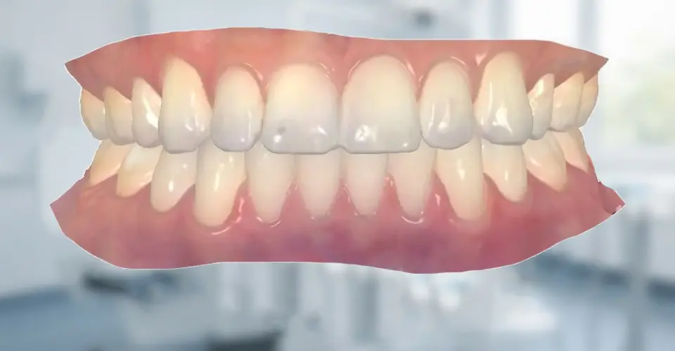
Improving Diagnosis, Planning & Surgical Precision
As a leader in advanced oral and maxillofacial care, our practice uses 3D imaging to improve diagnosis, treatment planning, and surgical precision. This technology provides detailed information about bone structure, tooth position, nerve pathways, sinus anatomy, and other important structures that cannot always be fully evaluated with traditional imaging. Having a complete understanding of your anatomy before treatment allows your oral surgeon to provide safer, more predictable care.
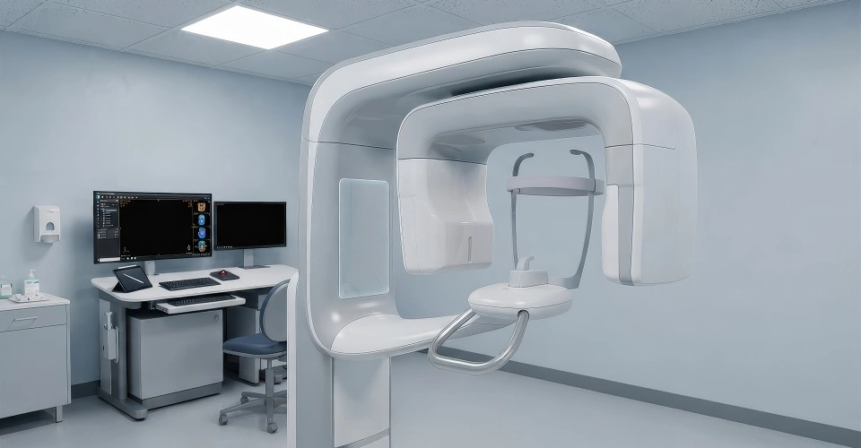
Enhancing Dental Implant Planning & All-on-X Restorations
One of the greatest benefits of 3D imaging is its ability to enhance dental implant planning. Successful dental implant placement requires careful evaluation of available bone volume, bone density, and the location of nearby anatomical structures such as nerves and sinuses. CBCT imaging allows your oral surgeon to determine the ideal implant position, evaluate whether bone grafting is needed, and create a customized surgical plan designed to maximize long-term implant success.
For patients receiving All-on-X dental implants or full-arch restorations, 3D imaging is an essential part of the treatment process. Advanced scans allow your surgeon to analyze the entire jaw, identify areas of stronger bone, and plan implant placement with exceptional accuracy. This technology supports guided implant surgery, helping improve efficiency, minimize surgical trauma, and provide patients with a more comfortable and predictable experience.
Wisdom Teeth, Facial Trauma & TMJ Evaluation
3D imaging also plays an important role in wisdom tooth evaluation and removal. Impacted wisdom teeth can develop near important nerves, blood vessels, and surrounding teeth. A CBCT scan allows your oral surgeon to clearly visualize the exact position of the tooth roots and their relationship to nearby structures. This detailed information helps reduce surgical risks and allows for a safer, more controlled procedure.
For patients experiencing facial trauma, 3D imaging provides invaluable information when diagnosing fractures and injuries involving the jaw, cheekbones, eye sockets, and other facial bones. Detailed three-dimensional views allow your oral surgeon to evaluate the severity and location of fractures, determine the best surgical approach, and restore facial structure and function with greater precision.
3D imaging is also extremely valuable in diagnosing and treating conditions involving the jaw joints, including temporomandibular joint (TMJ) disorders. The technology allows your surgeon to evaluate the bones and structures of the joint, identify abnormalities, and better understand the source of jaw pain, limited movement, or changes in function. This information helps guide treatment decisions and ensures that care is focused on addressing the underlying problem.
Oral Pathology, Patient Communication & Safety
When evaluating oral pathology, cysts, tumors, or other abnormalities, 3D imaging provides a detailed view of both the teeth and surrounding bone. This allows your oral surgeon to determine the size, location, and extent of a lesion while planning the safest approach for biopsy or surgical removal. Accurate imaging is an important step in diagnosing conditions early and providing appropriate treatment.
Another major advantage of CBCT imaging is improved patient communication. Because 3D scans create detailed visual models of your anatomy, your oral surgeon can clearly explain your diagnosis and treatment options. Seeing exactly what is happening beneath the surface allows patients to better understand their condition and feel more confident when making decisions about their care.
Modern 3D imaging systems are designed with patient safety in mind. Cone beam CT technology provides highly detailed images while using a focused beam that limits unnecessary radiation exposure compared to traditional medical CT scans. Your oral surgeon will only recommend imaging when it provides valuable diagnostic information that improves the safety, accuracy, or effectiveness of your treatment.
Personalized Care & An Investment in Excellence
By combining advanced imaging technology with specialized surgical training, oral and maxillofacial surgeons are able to provide more precise and personalized care than ever before. Whether you are considering dental implants, evaluating wisdom teeth, treating facial trauma, addressing TMJ concerns, or investigating oral pathology, 3D imaging provides the critical information needed for successful treatment planning.
Investing in advanced diagnostic technology is an important part of delivering exceptional oral surgery care. Through detailed 3D imaging, our team can better understand your unique anatomy, minimize uncertainty, and provide treatment that is safer, more comfortable, and more predictable. From diagnosis through recovery, this technology helps ensure that every aspect of your care is planned with precision and your long-term health in mind.